Specialized imaging for the detection of Deep Infiltrating Endometriosis (DIE) and Adenomyosis. We look deeper to find the answers to your pelvic pain.
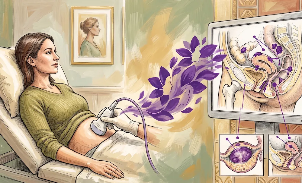
Standard pelvic ultrasounds frequently return “normal” results even when endometriosis is present. At Athena Women’s Ultrasound, our sonographers are specially trained in advanced endometriosis mapping. We actively look for nodules, scar tissue, and tethered organs that indicate Deep Infiltrating Endometriosis (DIE) and adenomyosis (endometriosis within the uterine muscle).
What We Evaluate During the Scan:
The Uterus and Ovaries: Checking for adenomyosis and endometriomas (chocolate cysts).
The Anterior Compartment: Evaluating the bladder wall for endometriotic nodules.
The Posterior Compartment: Checking the bowel, rectum, and the pouch of Douglas.
The “Sliding Sign”: A dynamic ultrasound technique used to see if the uterus and bowel slide freely against each other, or if they are stuck together by adhesions.
How to Prepare:
Because we evaluate the bowel during this scan, you may be required to take a mild bowel preparation (laxative) prior to your appointment to ensure clear imaging. Our reception team will provide specific instructions upon booking.
While we excel at detecting moderate to severe (DIE) endometriosis, superficial endometriosis may still only be definitively diagnosed via laparoscopic surgery.
Yes, please bring a referral from your GP or Gynaecologist specifically requesting an “Endometriosis Protocol” ultrasound. If you do not have a referral, do contact the clinic as we also offer GP Services.
Please contact the clinic directly to book an ultrasound appointment.
If you have a referral or anticipate needing an ultrasound in Perth, please reach out to our team to arrange your booking.